Image-based spatial gene expression
Transcriptomics Spatialhttps://www.biorxiv.org/content/10.1101/2023.12.13.571385v1
Image-based approaches, such as Vizgen Merscope, 10xGenomicx Xenium and Nanostring CosmX use microscopy and imaging technologies to visualize and quantify gene expression directly within tissue sections. They rely on fluorescently labeled probes which are designed to target specific mRNA sequences. Those approaches offer a high spatial resolution (subcellular), but for a limited number of targets (several hundreds, up to 5k for Xenium). The segmentation step is crucial to define the cell boundaries and to attribute a particular probe signal to a particular cell and obtain a gene x cell matrix associated to the spatial positions of each cell.
Production systems
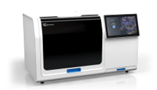
10xGenomics Xenium
Spatial transcriptomics approaches conserve the localization of transcript expression in tissue. This spatial information is crucial for contextualizing a transcriptomic phenotype observed at histological level and deducing its function, or for comparing pathology-induced expression variations within the same tissue. 10xGenomics Xenium is a state-of-the-art single cell spatial imaging platform which integrates high-resolution imaging and onboard data analysis enabling the processing of 400 mm² of tissue in <50 hrs (with nuclei-based segmentation). Xenium technology allows for the detection of gene pannels up to 5k targets, that can be customized.
See more about the technology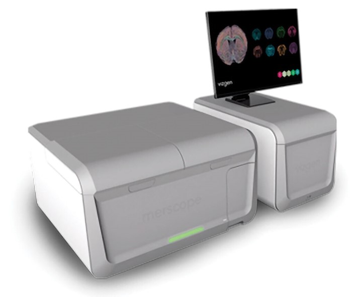
Vizgen Merscope
Vizgen Merscope is a spatial genomics platform that offers high-resolution gene expression mapping within tissues, enabling users to understand where and how genes are expressed. It features multiplexed imaging, allowing for the detection of up to thousands of RNA species, and provides subcellular resolution for detailed insights into the spatial organization within cells. Merscope allows for the design of custom gene panels tailored to specific research needs. Additionnaly, 6 proteins can be simultaneously detected within the same tissue.
See more about the technology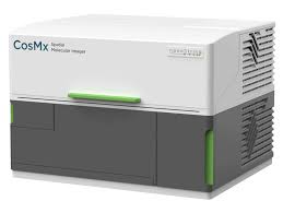
Nanostring Cosmx
CosMx SMI is the first high-plex in situ analysis platform to provide spatial multiomics with formalin-fixed paraffin-embedded (FFPE) and fresh frozen (FF) tissue samples at cellular and subcellular resolution. CosMx SMI enables rapid quantification and visualization of up to 6,000 RNA and 64 validated protein analytes. It is the flexible, spatial single-cell imaging platform that will drive deeper insights for cell atlasing, tissue phenotyping, cell-cell interactions, cellular processes, and biomarker discovery.
See more about the technologyAssociated pipelines
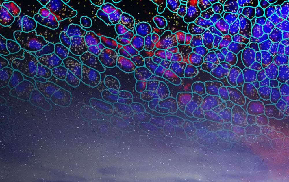
imaging-based in-situ spatial single-cell
This workflow integrates the key steps for a complete analysis of datasets generated either by Merscope, Xenium or Cosmx systems. It is based on the SpatialData and Anndata data structures from the scverse python ecosystem.
Go to pipeline documentation